The new scope came in Wednesday and I’d hoped to kick off this category with some great hi-res pics, but then was out sick on Thursday and wrapped up in meetings on Friday so I didn’t get a really good chance to play with it. I also still need to get a NIST traceable micro-ruler to calibrate it. If anyone knows of one for under $1200, I’d love to hear about it. Even without calibration though, I should be able to start posting some of the pics. At first glance, it looks pretty exciting. I can see the tiniest of scratches from our micro-abrasives really clearly, so with some luck and work, we should be able to learn a whole lot about all the stones and strops.
AWESOME!!!
cbw
Yes awesome. I am real anxious to start seeing some evidence of what we are actually doing at the sub micron level.
Phil
Can’t wait to see it in action! Thanks for making the investment Clay
Hi Clay,
I searched around and found a NIST-traceable stage micrometer for about $500, but I’m not sure it is good enough; I think it only goes down to 0.1mm (100 microns).
“Reticle Calibration Stage Micrometer, NIST traceable NT59-281 $495.00”
http://www.edmundoptics.com/microscopy/reticles-stage-micrometers/reticle-calibration-stage-micrometer/2826

Edmund Optics also has many other calibration targets, so you might also look around at:
http://www.edmundoptics.com/testing-targets/test-targets/image-analysis-test-targets/
http://www.edmundoptics.com/microscopy/reticles-stage-micrometers/
I also found this company that sells stage micrometers with NIST-traceable certificates. It seems if you buy the stage-micrometer from them, they lower the price for NIST-traceable calibration.
http://www.aig-imaging.com/NIST-Traceable-Calibration.html
http://www.aig-imaging.com/Stage-Micrometer-Scales.html
Other places I looked at had fancier stage micrometers, but were very expensive ($1200+ for NIST-traceable):
http://www.emsdiasum.com/microscopy/products/magnifier/stage.aspx
http://www.tedpella.com/calibrat_html/calib.htm
Sincerely,
–Lagrangian
P.S. Another possibility is to get a mechanical microscope stage with micrometers. That is, it is a stage that can move in the X, or X-and-Y directions where each direction is literally moved by a micrometer-head. Presumably, if the micrometer-heads are NIST certified, then the microscope stage is very accurate. It may be expensive, so don’t know if it would cost less. And I don’t know if they actually are avaiable as NIST-traceable.
http://www.edmundoptics.com/microscopy/microscopy-mechanics/linear-positioning-stages/30mm-single-axis-crossed-roller-translation-stages/3216

I have been watching this thread for a while. Just curious why do you feel that you need a traceable standard? That will just add tremendous cost, but I do not see any benifit for you / us? Tell me exactly what you are trying to accomplish here, and I will make an inquiry with a friend who is in the optics measuring business (adcole inc.) as I think there are several work arounds for this if you can take a reference that is not “traceable”
[quote quote=“AnthonyYan” post=3660]..
P.S. Another possibility is to get a mechanical microscope stage with micrometers. That is, it is a stage that can move in the X, or X-and-Y directions where each direction…etc [/quote]
Yes I agree with Anthony here. In fact you are going to need this to handle even using you new device, as you certainly can not span and observe any item in such a magnified field of view without assistance from an axis controller of some sort.
unsure what they use but they test our knifes @ work
Starting to have some fun with the new scope ![]() The stage micrometer came in and I’ve calibrated it at 800x. I had to send the 100x objective lens back as it was defective, so I can’t get up to 2000x yet… So far, the images I’m getting at 800x are great and I’m learning a lot in a short amount of time. I can’t wait to share what I’m learning with you guys, just need more hours in the day!
The stage micrometer came in and I’ve calibrated it at 800x. I had to send the 100x objective lens back as it was defective, so I can’t get up to 2000x yet… So far, the images I’m getting at 800x are great and I’m learning a lot in a short amount of time. I can’t wait to share what I’m learning with you guys, just need more hours in the day!
Hi Clay,
Sounds exciting! ![]()
I’m curious… which stage micrometer did you end up getting?
Sincerely,
–Lagrangian
[quote quote=“AnthonyYan” post=4057]Hi Clay,
Sounds exciting! ![]()
I’m curious… which stage micrometer did you end up getting?
Sincerely,
–Lagrangian[/quote]
hey Anthony, help me out here. .. I will differ here to what you tell me: I seem to recall, from many many years past when sleeping through one my physics classes that there was some professor droning on about nanometers, and the wavelength of (human) visiable light and 1/2 a micron and, well anyway, 2000X? optical?? I am a bit lost here…
Hey Lagrangian,
I ended up with one from Ted Pella, Inc. It is: “Micrometer Scale-Reflected Light 2mm in .01mm divisions” It’s probably not ready for NASA, but I think it is good enough for the kind of work I’m doing. The microscope stage has built in axis control with pretty fine adjustments, so I didn’t need that feature - I can easily scroll along the length of the blade or from shoulder to edge which is good enough for me. Next I need to design and build a stage that will hold my blades so I can take the samples in and out quickly without a lot of fiddling.
-Clay
[quote quote=“AnthonyYan” post=4057]Hi Clay,
Sounds exciting! ![]()
I’m curious… which stage micrometer did you end up getting?
Sincerely,
–Lagrangian[/quote]
Hi Clay:
Thanks! ![]()
Hi BassLakeDan:
For a variety of reasons, visible wavelength is often discussed in terms of nanometers, and sometimes in angstroms. But it’s just all unit conversions by factors of ten.
Visible light is approximately in the range of 0.4 to 0.7 microns (okay, if you are really picky, the 0.38 to 0.74 microns). If you want, that’s 400 to 700 nanometers, or 4000 to 7000 angstroms.
https://en.wikipedia.org/wiki/Visible_light
Optical microscopes use visible light to image stuff, so they’re pretty good until you start getting down to sub-wavelength features. Below about half a wavelength, you will probably not see anything. Current high-end optical microscopes have a resolution of about 0.2 microns. You can get better than this optically, but doing so requires extremely fancy and high-tech methods. So it’s only in a research lab that you would be likely to see an optical microscope with better than 0.2 micron resolution.
A ton of really good microscope information can be found here, on Nikon’s microscope web page:
http://microscopyu.com/
To get to higher resolutions, one can use non-optical methods, such as using light outside of the visibel spectrum, or things like electron-microscopes, atomic-force microscopes, and scanning-tunnelling-electron micrscopes.
Sincerely,
–Lagrangian
P.S. btw, as a knife and sharpening enthusiast, I’m conceptually anchored at 0.5 microns because the sharpness of a modern razor is about 0.4 microns (according to Prof. John D. Verhoeven), and visible light is around 0.4 to 0.7 microns. So whenever I think about knife edges, microscope pictures, and the size of abrasive particles, I’m thinking in terms of 0.5 microns. This is why I think it’s cool that Clay got a stage micrometer… because in any photos he shows, we will be able to have some idea of the width of scratches in comparison to edge sharpness and abrasive grits.
P.P.S. I’ve only been a knife enthusiast for about a year, but over that time I’ve collected a short list of “length-scales” related to knives. It is kind of like a time-line, but instead of time, the axis is length (microns). Here’s a partial list:
50-100 microns = Approximate diameter of human hair (varies hugely; this is only part of the range)
25.4 microns = 0.001 inches (1 mil). Standard resolution for an imperial caliper
2.54 microns = 0.0001 inches (0.1 mil). Standard resolution for an imperial micrometer.
0.36-0.74 microns = Wavelength of visible light
0.4 microns = Sharpness of a modern razor blade.
0.2 microns = resolution limit of optical microscopes
0.05 microns = Sharpness of diamond coated razors.
0.005 microns = Sharpness of a diamond microtome knife.
0.003 microns = Sharpness of concoidally fractured obsidian.
0.00034 microns = van der Waals diameter of a carbon atom.
Atomic-force microscopes (AFM) and scanning-tunneling-electron-microscopes (STEM) use needles which are atomically sharp; they literally have a single atom at the tip.
Sources:
https://en.wikipedia.org/wiki/Hair#Description
https://en.wikipedia.org/wiki/Visible_light
http://www-archive.mse.iastate.edu/fileadmin/www.mse.iastate.edu/static/files/verhoeven/KnifeShExps.pdf
http://microscopyu.com/articles/optics/index.html
http://www.technologyreview.com/computing/25988/
http://www.tedpella.com/diamond_html/diamondk.htm
http://en.wikipedia.org/wiki/Obsidian
https://en.wikipedia.org/wiki/Van_der_Waals_radius
P.P.P.S. If you are interested in the conversion between grit numbers and microns, then have a look at Komitadjie’s Grand Unified Grit Chart. Here are some plots by Mr. Wizard using the data that Komitadjie has compiled:
http://www.knifeforums.com/forums/showpost.php?post/2387907/

thanks Anthony for all the great info.. i will wait to see Clay photos.. I am sort of a “proof of the pudding is in the tasting” type so.. if Clay can bring this off then he probably can get ‘on the side’ contract work for various labs that need those persons skilled in the use of scopes at these ranges. What he is up to is no simple task, and the hurdles to be overcome should not be under estimated. But, of course I want to see the results as much as anyone, so I wish all 100% success on the project !!!
I just saw this post from Magnaminous_G on BladeForums.com:
http://www.bladeforums.com/forums/showthread.php/975577-This-might-interest-serious-sharpeners-free-SEM-imaging
[quote quote=“Magnaminous_G of www.BladeForums.com”]This might interest serious sharpeners - free SEM imaging
It looks like ASPEX is now offering free SEM imagery of whatever you send to them (limit of 2 samples). It might be really cool to see a very refined edge under a SEM.
http://www.aspexcorp.com/Resources/SendUsYourSample.aspx[/quote]

If they are able to take SEM pictures looking directly into the edge (the same as Prof. John D. Verhoeven), then they may be able to measure the actual sharpness of your knife edge. That would be amazingly cool… With modern ultra-fine abrasives, do you think your edge is sharper than 0.4 microns? Now you can find out!
http://www-archive.mse.iastate.edu/fileadmin/www.mse.iastate.edu/static/files/verhoeven/KnifeShExps.pdf
Sincerely,
–Lagrangian
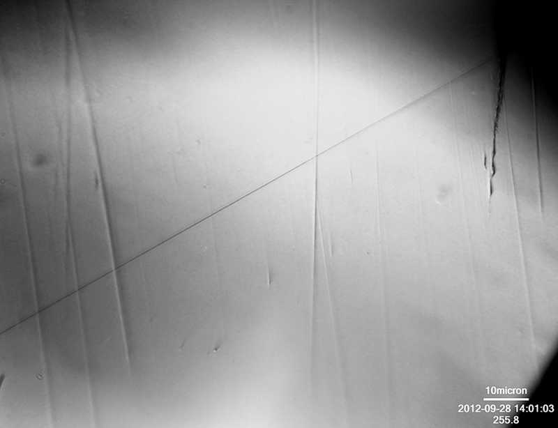
First image at 2000x and only a few seconds in which to post it:
The long diagonal scratch measures between .15um and .20um. I will have to work on making the annotations and measurements show up more on Monday, but for now, I’m really excited that I’ve got it operable at last.
Attachments:
Hi Clay,
Wow, very cool!
btw, I’m not a microscopy expert. But as a physics major, I’m not sure that you can reliably measure features smaller than about 0.2 microns.
The resolution limit of optical microscopes is around 0.2 microns (which is 1/2 wavelength of visible light which is 0.4-0.7 microns). A conventional microscope cannot resolve smaller features. It’s a limitation of the physics of light. Basically, light is a wave, and so it will diffract. Light diffracts slightly when going through a microscope objective, and that diffraction causes some blurring that cannot be removed by any standard optics.
There are some non-standard optics which can get past this resolution limit, but they are very fancy, very expensive, and very technical. Computer chip manufacturers use some of these super-fancy techniques to optically stencil the patterns for transistors etc. And fluorescent microscopy can do some interesting stuff. Also, there is high-end research into microscope resolution. But all that aside, it seems very difficult to get to 0.2 micron resolution not to mention finer resolutions. The actual theoretical limit can be finer than this, but I have heard that 0.2 microns is basically the practical limit.
Some references:
http://en.wikipedia.org/wiki/Diffraction-limited_system
http://www.microscopyu.com/articles/optics/mtfintro.html
I very much like Nikon’s webpage on general microscopy:
http://www.microscopyu.com/articles/optics/index.html
If you know someone who is more deeply familiar with the technical aspects of microscopy, maybe you can ask them? Or maybe you can ask the manufacturer of your microscope too.
Sincerely,
–Lagrangian
P.S. For those of you with a technical background: The diffraction basically causes the “true image” to be convolved with an Airy function. Conceptually, this is simlar to applying a Gaussian Blur in Adobe Photoshop (which is also a convolution). Using Fourier analysis, one can look in the frequency domain, where this is like applying a low-pass filter, after which, all the high-frequency details cannot be recovered. Some of the semi-technical details for this are discussed in Nikon’s webpage in the links above. For more technical details, one can go to undergraduate physics textbook on wave-mechanics, or a textbook on optics.
Lagrangian,
Thanks again for the informative post! I’m certainly no expert in microscopy either but I’ve read a few things here and there that agree with what you’ve said, especially about the limits of what can be achieved with optical scopes. I think you can see features below .2um but they are fuzzy at best, you just get the hint of them and I’m sure you’re right that you can’t state measurements in that range with confidence. For my purposes, I’m pretty content with this level for now as it at least helps me visualize more clearly what’s going on with some of the finer grits. It’s also helpful for analyzing different techniques like edge-leading vs edge-trailing. Once of these days, I’ll have to scheme regular access to and SEM ![]()
While that would be interesting, I think you may get beyond what you’re trying to “see” (from a practical stand point) Looking at the surface of polished steel where the grain structure looks like the surface of the moon may not actually be helpful in terms of sharpness. It would be interesting to figure out how much is enough, though.
For me, I find other elements like focus and depth of field to be more helpful than magnification.
Ken
Hey Ken,
My interest in the higher magnifications is to better understand what is happening with super fine polishing grits and substrates e.g. cow leather vs kangaroo vs horse vs nano-cloth etc… The studies I’ve already done have told me a lot, but I want to keep learning. I also want to be able to view and measure the edge end-on after it’s been sharpened with various media.
While that would be interesting, I think you may get beyond what you’re trying to “see” (from a practical stand point) Looking at the surface of polished steel where the grain structure looks like the surface of the moon may not actually be helpful in terms of sharpness. It would be interesting to figure out how much is enough, though.
For me, I find other elements like focus and depth of field to be more important than magnification.
Ken[/quote]